SIGN UP FOR NEWSLETTER
Signup for our newsletter to get notified about sales and new products

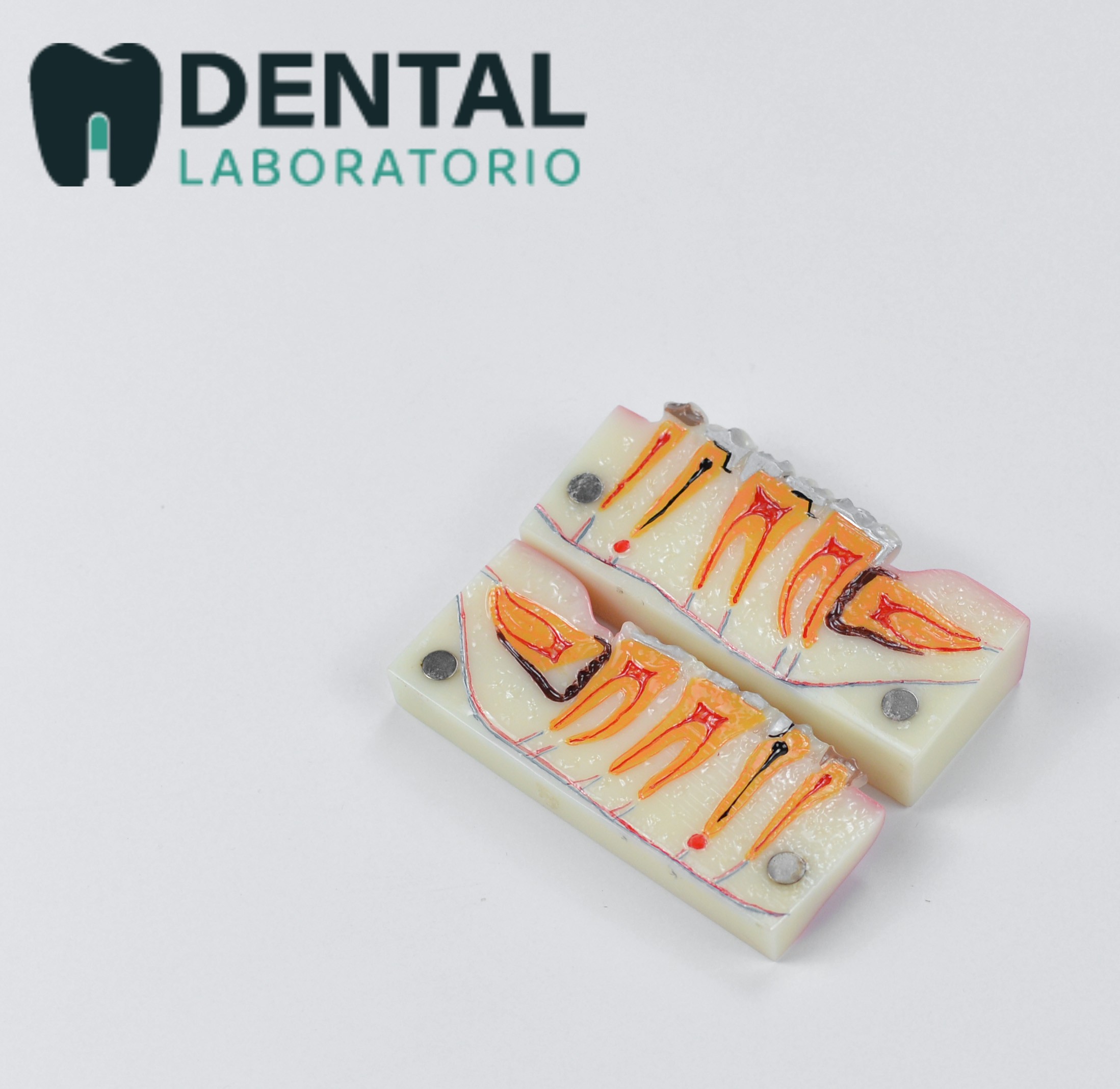


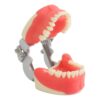
Pathological posterior tooth model anatomy, 4 times magnification.
The detachable, magnetic adsorption design makes the model easy to be demonstrated Inner tooth structure.
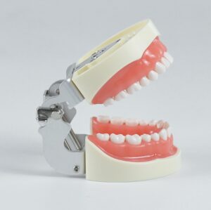
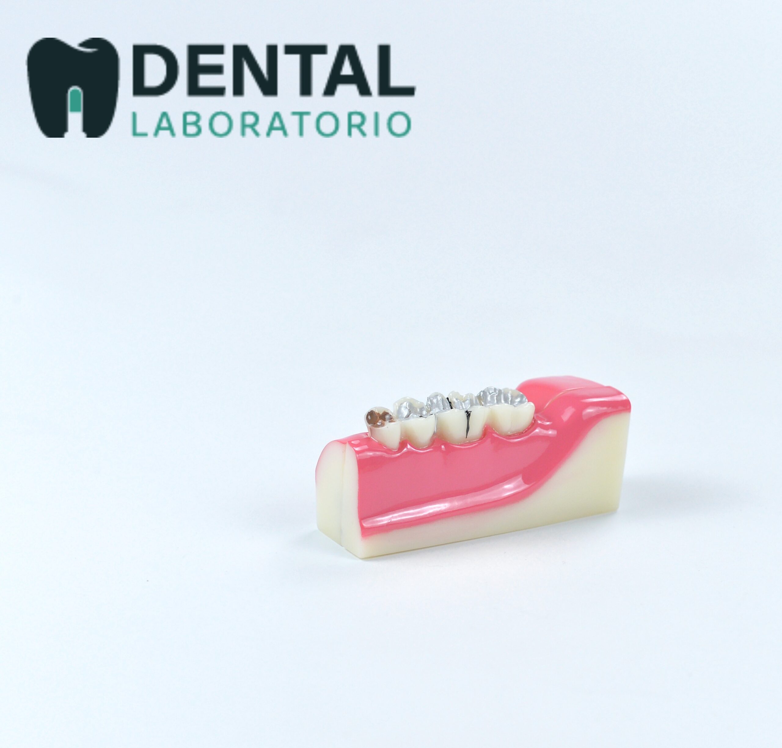
A good dentist simulation tool for better patient education.
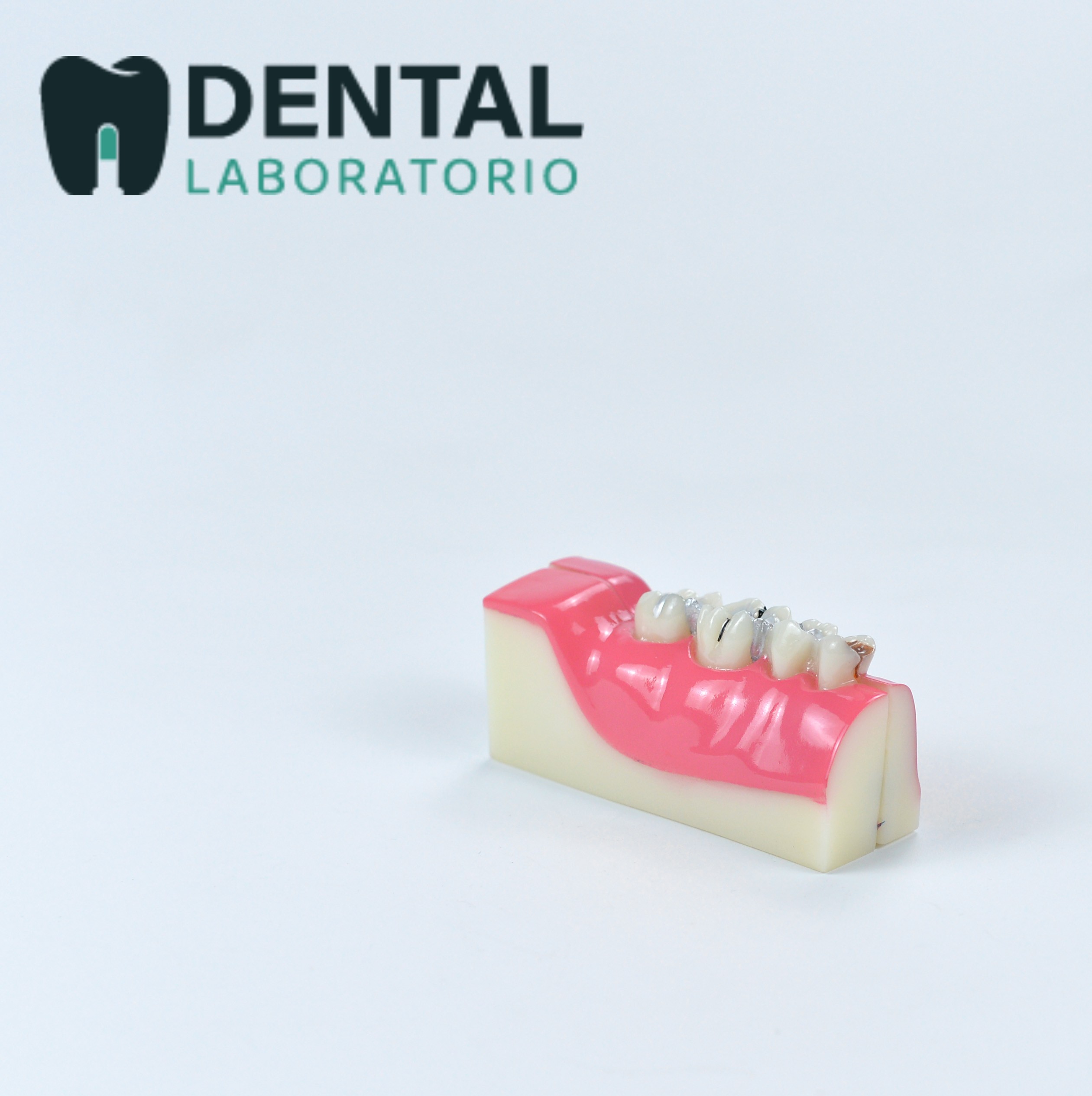
Dimension: 10.5*3.2*4.3 cm.
Free shipping.
Deal of the Day
Original price was: $40.00.$34.00Current price is: $34.00.
Want to buy even more quantity?
Wholesale Price Inquiry
This tooth model anatomy is an effective tool for patient education. It displays a posterior tooth affected by caries, root canal pulp, nerves, a horizontal inclination wisdom tooth, and apical cysts.
The model is composed of five teeth embedded in a red gingival layer that can be separated into two parts, allowing for a four-time magnification of the internal tooth structure.
This model provides a detailed and realistic representation of the anatomy of a tooth, enabling patients to better understand the diagnosis and treatment plan that is best suited for them.
| Weight | 1 kg |
|---|---|
| Dimensions | 10 × 10 × 7 cm |